
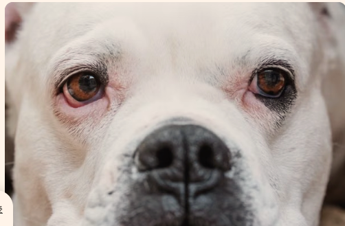
Dog eye photo · No clinical history provided
Photo-based observation
Visible pink lower eyelid tissue exposure
The photo shows exposed pink tissue along the lower eyelid margins, more noticeably prominent in image left. Because unexplained eye redness or eyelid margin exposure can stem from anatomical shape or surface irritation, contact a veterinarian today or within 24 hours to determine if an exam is needed.
When to contact a veterinarian
Contact a veterinarian today or within 24 hours
Unexplained ocular redness or exposed eyelid lining warrants prompt veterinary guidance to rule out underlying irritation or corneal damage.
A visible pattern is not a diagnosis. A photo cannot confirm that waiting is safe.
What is visible in the photo
Exposed lower eyelid tissue
Pink inner eyelid lining is visible along both lower lids, appearing noticeably more exposed and drooping on image left compared to image right.
Image detail: clearly visible
Possible explanations
These are possibilities to discuss with a veterinarian, not confirmed conditions.
Conformational eyelid laxity
Loose lower eyelid margins (ectropion) can naturally expose normal pink conjunctiva, but conformation cannot be distinguished from active irritation without clinical evaluation.
Conjunctival irritation or inflammation
Allergens, foreign material, or mild infection can cause visible redness and swelling of the eyelid lining, though specific causes require an in-person eye exam to detect.
What to do next
- Contact your veterinarian today or within 24 hours to schedule an evaluation or discuss next steps.
- Prevent your dog from rubbing or pawing at the face to protect the eyes from secondary injury.
- Do not administer any human medications, over-the-counter drops, or ointments into the eyes.
Signs that need prompt care
- Seek immediate veterinary care for severe eye pain, sudden vision changes, trauma, chemical exposure, or marked cloudiness.
- Seek same-day assessment if you notice squinting, an eye held closed, repeated pawing, or new yellow or green discharge.
What this photo cannot tell us
- A single still photo cannot assess or exclude a corneal scratch, tear-film deficiency, or elevated eye pressure.
- The image cannot reveal duration, history of rubbing, or whether the exposure is new or lifelong.
Details that would help
- Has this appearance been present long-term, or did it develop recently?
- Have you seen any pawing, rubbing, squinting, or signs of pain?
- Have you noticed any watery, mucus, or colored discharge from either eye?
Your Vet Visit Prep Sheet
- Note whether the redness or eyelid droop worsens outdoors or at certain times of day.
Questions for your veterinarian
- Is this eyelid shape normal conformation for my dog, or is there an active inflammatory condition?
- Does my dog need a fluorescein stain to check for corneal scratches or an eye pressure check?